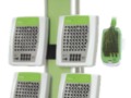
Grael 4K PSG/EEG Specifications Sheet
The post Grael 4K PSG/EEG Specifications Sheet appeared first on Compumedics Neuroscan.
Grael 4K PSG/EEG Specifications Sheet
The post Grael 4K PSG/EEG Specifications Sheet appeared first on Compumedics Neuroscan.
The CURRY NeuroImaging platform and MEG have a history stretching back over 25 years. CURRY was first conceived as a product in the early 1990’s when Philips Electronics investigated the feasibility of developing it’s own MEG hardware platform. Ultimately, the hardware platform did not survive, but the software, along with its core engineering architects, Dr. Manfred Fuchs & Dr. Michael Wagner, continued on. When Philips exited the MEG business, CURRY and the development team were purchased by Neuroscan. At this time, the UNIX-based CURRY platform appealed more to the research community than to the clinical market. By 1999, publications were emerging describing the application of CURRY for cortical localization of EEG and MEG activity for tactile and auditory sensory input. However, “novel developments” and “new approaches to detailed localization of specific epileptic discharges” as well as identification of functionally critical areas of the brain controlling language and memory using CURRY, were also being reported in the clinical literature. Migration of CURRY from the UNIX to Windows platform in 2003 resulted in a rapid expansion of the use of CURRY in both the research and clinical worlds.
The benefits associated with CURRY’s ability to integrate MEG with EEG and co-register both kinds of high temporal resolution functional imaging data with the structural neuroimaging data including MRI, CT, DTI, as well as PET, SPECT and fMRI accelerated the the adoption of the software by both the research and clinical communities. Early clinical adopters, such as Dr. John Ebersole, supported and championed the benefits of source localization tools such as CURRY, contributing to the development of specific source analysis billing codes for EEG and MEG. For a long time, CURRY has been the de-facto software platform for clinical MEG community, particularly for those assessing epilepsy. This has culminated in the adoption of CURRY as the standard analysis platform by the European E-pilepsy Consortium.
For the CURRY team, integrating CURRY with the KRISS MEG hardware represents a full circle of development. With long-term future development plans for both hardware and software, CURRY MEG will offer an expanding list of benefits based on the first fully integrated platform combining EEG, MEG, multi-modal neuroimage co-registration and source reconstruction from a single provider – Compumedics Neuroscan.
The post Magnetoencephaloghy (MEG) and CURRY – A long history together appeared first on Compumedics Neuroscan.
A Comprehensive Epilepsy Solution In a Single Seamless Platform. Compumedics and Neuroscan products are emerging as the standard in facilities who require a single data acquisition and analysis system that can combine the best of clinical neurology and neuroscience, as well as the premiere hybrid of both disciplines. We continue to strive toward advanced products that offer no compromises, whether applied in the research laboratory or in clinical settings.
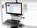
Neuvo LTM
The Ultimate Long-term EEG Monitoring System.
From 64 to, now, 512 channels of EEG, the Neuvo LTM system exceeds all your previous expectations. Born from and integrated with the Neuroscan Brain Research technologies, Neuvo provides that seamless transition for clinical neuroscience.
Profusion 4 EEG
Unlimited potential in routine and LTM EEG.
An advanced user-friendly interface provides the user with reports, mapping, template matching and multiple high resolution video windows. Open your clinical EEG files in Curry with a single click. Functions with all Compumedics EEG amplifiers.
Profusion Nexus
Complete laboratory management system of patient data and laboratory control for class leading efficiency. Includes management of appointments, waitlists, process workflow, disk space and laboratory resources.
Grael-HD EEG System
High definition EEG for the Clinical World.
Reliable and flexible EEG studies for the office, clinic or hospital. The Grael EEG system is ideal for clinical and LTM EEG.
[contact-form-7]
The post Compumedics Neuroscan Neurodiagnostics appeared first on Compumedics Neuroscan.
Huang J, Colrain IM, Melendres MC, Karamessinis LR, Pepe ME, Samuel JM, Abi-Raad RF, Trescher WH, Marcus CL. Cortical processing of respiratory afferent stimuli during sleep in children with the obstructive sleep apnea syndrome. Sleep. 2008;31(3):403–10.
Melendres MC, Marcus CL, Abi-Raad RF, Trescher WH, Lutz JM, Colrain IM. Respiratory-related evoked potentials during sleep in children. Sleep. 2008;31(1):55–61.
Abi-Raad R, Tan WKM, Bennet L, Gunn AJ, Davis SL, Gluckman PD, Johnston BM and Williams CE Role of the Cerebrovascular and Metabolic Responses in the Delayed Phases of Injury after Transient Cerebral Ischemia in Fetal Sheep. Stroke 1999. 30(12):2735–2741.
Höper J, Kessler M, Abi-Raad R, Funk R Oxygen-dependent Regulation of Capillary Flow. Fact or Fiction? Advances in Experimental Medicine & Biology 1997. 428:415–24.
Harrison DK, Abi-Raad R, Newton DJ, McCollum PT Transcutaneous H2 Clearance — A New Least-invasive Method for Assessing Skin Blood Flow. Advances in Experimental Medicine & Biology 1994. 361:181–6.
Harrison DK, Abi-Raad R, Newton DJ and McCollum PT Transcutaneous Hydrogen Clearance — A New Non-invasive Technique for Assessing Blood Flow in Human Skin. Physiol Meas 1994 15(1): 89–100.
Fuchs M, Ford MR, Sands S, Lew HL. Overview of dipole source localization. Phys Med Rehabil Clin N Am. 2004 Feb;15(1):251–62.
Ford MR, Sands S, Lew HL. Overview of artifact reduction and removal in evoked potential and event-related potential recordings. Phys Med Rehabil Clin N Am. 2004 Feb;15(1):1–17.
Ford M.R.; Sidman R.D.; Khalil M.A.; Lan K. Cortical potential differences in mild traumatic brain injury for the AEP P300 component Electroencephalography and Clinical Neurophysiology, 1997 102(1) , 21P-21P.
Ford, Martin R. Khalil, Mohamed. Evoked Potential Findings in Mild Traumatic Brain Injury 1: Middle Latency Component Augmentation and Cognitive Component Attenuation. Journal of Head Trauma Rehabilitation. 1996 11(3):1–15.
Ford, Martin R; Khalil, Mohamed Evoked Potential Findings in Mild Traumatic Brain Injury 2: Scoring System and Individual Discrimination. Journal of Head Trauma Rehabilitation. 1996 11(3):16–21.
Sidman RD, Ford MR, Ramsey G. Optimal electrode placements for adequate spatial sampling of auditory evoked potentials. Brain Topogr. 1994 Spring;6(3):227–30.
Ford MR, Sidman RD, Ramsey G. Spatio-temporal progression of the AEP P300 component using the cortical imaging technique. Brain Topogr. 1993 Fall;6(1):43–50.
Sidman R. D. ; Major D. J. ; Ford M. R. ; Ramsey G. G. ; Schlichting C. Age-related features of the resting pattern-Reversal visual evoked response using the dipole localization method and cortical imaging technique. J Neurosci Methods. 1991, vol. 37, no1, pp. 27–36
Sidman RD, Ford MR, Ramsey G, Schlichting C. Age-related features of the resting and P300 auditory evoked responses using the dipole localization method and cortical imaging technique. J Neurosci Methods. 1990 Jul;33(1):23–32.
Sidman RD, Kearfott RB, Major DJ, Ford MR, Hill CD, Smith DB, Lee L, Kramer R. Development and application of mathematical techniques for the non-invasive localization of the sources of scalp-recorded electric potentials. In Biomedical Modelling and Simulation, Eisenfeld J & Levine DS (Eds.) JC Baltzer AG, Scientific Publishing Co., 1989: 133–157.
Glueck BC, Ford MR, Molyn MA. Computer analysis of the electroencephalogram. Psychiatric Annals 1988 18:236–245.
Ford MR, Goethe JW, Dekker DK. EEG coherence and power in the discrimination of psychiatric disorders and medication effects. Biol Psychiatry. 1986 Oct;21(12):1175–88.
Ford MR, Goethe JW, Dekker DK. EEG coherence and power changes during a continuous movement task. Int J Psychophysiol. 1986 Jul;4(2):99–110.
Ford MR. Interpersonal stress and style as predictors of short– and long-term outcome. Biofeedback Self Regul. 1985 10:223–239.
Ford MR, Stroebel CF, Strong P, Szarek BL. Quieting response training: predictors of long-term outcome. Biofeedback Self Regul. 1983 Sep;8(3):393–408.
Ford MR, Stroebel CF, Strong P, Szarek BL. Quieting response training: long-term evaluation of a clinical biofeedback practice. Biofeedback Self Regul. 1983 Jun;8(2):265–78.
Ford MR, Stroebel CF, Strong P, Szarek BL. Quieting response training: treatment of psychophysiological disorders in psychiatric inpatients. Biofeedback Self Regul. 1982 Sep;7(3):331–9.
Ford MR. Biofeedback treatment for headaches, Raynaud’s disease, essential hypertension and irritable bowel syndrome: A review of the long-term, follow-up literature. Biofeedback Self Regul. 1982 Sep;7(4):521–536.
Mirabile CS, Ford MR. A clinically useful polling technique for establishing susceptibility to motion sickness. Perceptual and Motor Skills. 1982 54:987–991.
Stroebel CF, Ford MR. Biofeedback – Quieting response treatment of primary Raynaud’s disease. In Biofeedback – Basic Problems and Clinical Applications, Richter-Heinrich E & Miller NE (Eds), VEB Deutscher Verlag de Wissenschaften, Berlin, 1982.
Ford M, Bird BL, Newton FA, Sheer D. Maintenance and generalization of 40-Hz EEG biofeedback effects. Biofeedback Self Regul. 1980 Jun;5(2):193–205.
Spydell JD, Ford MR, Sheer DE. Task dependent cerebral lateralization of the 40 Hertz EEG rhythm. Psychophysiology. 1979 Jul;16(4):347–50.
Bird BL, Newton FA, Sheer DE, Ford M. Behavioral and electroencephalographic correlates of 40-Hz EEG biofeedback training in humans. Biofeedback Self Regul. 1978 Mar;3(1):13–28.
Bird BL, Newton FA, Sheer DE, Ford M. Biofeedback training of 40-Hz EEG in humans. Biofeedback Self Regul. 1978 Mar;3(1):1–11.
Spydell JD, Ford MR, Sheer DE. Task dependent cerebral lateralization of the 40 Hertz EEG rhythm. Psychophysiology. 1979 Jul;16(4):347–50.
Plummer C, Wagner M, Fuchs M, Vogrin S, Litewka L, Farish S, Bailey C, Harvey AS, Cook MJ. Clinical utility of distributed source modelling of interictal scalp EEG in focal epilepsy. Clin Neurophys. 2010, 121(10) , 1726–1739.
Plummer C, Wagner M, Fuchs M, Harvey AS, Cook MJ. Dipole Versus Distributed EEG Source Localization for Single Versus Averaged Spikes in Focal Epilepsy J Clin Neurophysiol. 2010, 27(3):141–162.
Fuchs M, Wagner M, Kastner J. Development of volume conductor and source models to localize epileptic foci. J Clin Neurophysiol. 2007 Apr;24(2):101–19.
Wagner M, Fuchs M, Kastner J. Evaluation of sLORETA in the presence of noise and multiple sources. Brain Topogr. 2004;16(4):277–80.
Fuchs M, Wagner M, Kastner J. Confidence limits of dipole source reconstruction results. Clin Neurophysiol. 2004 Jun;115(6):1442–51.
Fuchs M, Ford MR, Sands S, Lew HL. Overview of dipole source localization. Phys Med Rehabil Clin N Am. 2004 Feb;15(1):251–62.
Fuchs M, Kastner J, Wagner M, Hawes S, Ebersole JS. A standardized boundary element method volume conductor model. Clin Neurophysiol. 2002 May;113(5):702–12.
Fuchs M, Wagner M, Kastner J. Boundary element method volume conductor models for EEG source reconstruction. Clin Neurophysiol. 2001 Aug;112(8):1400–7.
Schmitt U, Louis AK, Darvas F, Buchner H, Fuchs M. Numerical aspects of spatio-temporal current density reconstruction from EEG-/MEG-data. IEEE Trans Med Imaging. 2001 Apr;20(4):314–24.
Darvas F, Schmitt U, Louis AK, Fuchs M, Knoll G, Buchner H. Spatio-temporal current density reconstruction (stCDR) from EEG/MEG-data. Brain Topogr. 2001 Spring;13(3):195–207.
Waberski TD, Gobbelé R, Herrendorf G, Steinhoff BJ, Kolle R, Fuchs M, Paulus W, Buchner H. Source reconstruction of mesial-temporal epileptiform activity: comparison of inverse techniques. Epilepsia. 2000 Dec;41(12):1574–83
Buchner H, Gobbele R, Waberski TD, Wagner M, Fuchs M. Evidence for independent thalamic and cortical sources involved in the generation of the visual 40 Hz response in humans. Neurosci Lett. 1999 Jul 9;269(2):59–62.
Fuchs M, Wagner M, Köhler T, Wischmann HA. Linear and nonlinear current density reconstructions. J Clin Neurophysiol. 1999 May;16(3):267–95.
Ossenblok P, Fuchs M, Velis DN, Veltman E, Pijn JP, da Silva FH. Source analysis of lesional frontal-lobe epilepsy. IEEE Eng Med Biol Mag. 1999 May-Jun;18(3):67–77.
Fuchs M, Wagner M, Wischmann HA, Köhler T, Theissen A, Drenckhahn R, Buchner H. Improving source reconstructions by combining bioelectric and biomagnetic data. Electroencephalogr Clin Neurophysiol. 1998 Aug;107(2):93–111.
Fuchs M, Drenckhahn R, Wischmann HA, Wagner M. An improved boundary element method for realistic volume-conductor modeling. IEEE Trans Biomed Eng. 1998 Aug;45(8):980–97.
Waberski TD, Buchner H, Lehnertz K, Hufnagel A, Fuchs M, Beckmann R, Rienäcker A. Properties of advanced headmodelling and source reconstruction for the localization of epileptiform activity. Brain Topogr. 1998 Summer;10(4):283–90.
Buchner H, Gobbelé R, Wagner M, Fuchs M, Waberski TD, Beckmann R. Fast visual evoked potential input into human area V5. Neuroreport. 1997 Jul 28;8(11):2419–22.
Buchner H, Knoll G, Fuchs M, Rienäcker A, Beckmann R, Wagner M, Silny J, Pesch J. Inverse localization of electric dipole current sources in finite element models of the human head. Electroencephalogr Clin Neurophysiol. 1997 Apr;102(4):267–78.
Buchner H, Waberski TD, Fuchs M, Wischmann HA, Wagner M, Drenckhahn R. Comparison of realistically shaped boundary-element and spherical head models in source localization of early somatosensory evoked potentials. Brain Topogr. 1995 Winter;8(2):137–43.
Buchner H, Waberski TD, Fuchs M, Wischmann HA, Beckmann R, Rienäcker A. Origin of P16 median nerve SEP component identified by dipole source analysis–subthalamic or within the thalamo-cortical radiation? Exp Brain Res. 1995;104(3):511–8.
Buchner H, Fuchs M, Wischmann HA, Dössel O, Ludwig I, Knepper A, Berg P. Source analysis of median nerve and finger stimulated somatosensory evoked potentials: multichannel simultaneous recording of electric and magnetic fields combined with 3D-MR tomography. Brain Topogr. 1994 Summer;6(4):299–310.
Fuchs M, Wagner M, Kastner J. Development of volume conductor and source models to localize epileptic foci. J Clin Neurophysiol. 2007 Apr;24(2):101–19.
Wagner M, Fuchs M, Kastner J. Evaluation of sLORETA in the presence of noise and multiple sources. Brain Topogr. 2004;16(4):277–80.
Fuchs M, Wagner M, Kastner J. Confidence limits of dipole source reconstruction results. Clin Neurophysiol. 2004 Jun;115(6):1442–51.
Fuchs M, Kastner J, Wagner M, Hawes S, Ebersole JS. A standardized boundary element method volume conductor model. Clin Neurophysiol. 2002 May;113(5):702–12.
Fuchs M, Wagner M, Kastner J. Boundary element method volume conductor models for EEG source reconstruction. Clin Neurophysiol. 2001 Aug;112(8):1400–7.
Näätänen R, Kujala T, Kreegipuu K, Carlson S, Escera C, Baldeweg T, Ponton C. The mismatch negativity: an index of cognitive decline in neuropsychiatric and neurological diseases and in ageing. Brain. 2011.
Politte, D. Prior, F. Ponton, C. Nolan, T. Larson-Prior, L. Sources of non-physiologic noise in simultaneous EEG-fMRI data: A phantom study. Conf Proc IEEE Eng Med Biol Soc. 2010;1:5129–32.
Ponton CW, Bernstein LE, Auer ET. Mismatch negativity with visual-only and audiovisual speech. Brain Topogr. 2009; 21(3–4):207–215.
Bernstein LE, Auer ET, Wagner M, Ponton CW. Spatiotemporal dynamics of audiovisual speech processing. NeuroImage, Neuroimage. 2008; 39(1):423–35.
Shafer VL, Ponton CW, Datta H, Mora M, Schwartz RG. Neurophysiological Indices of Attention to Speech in Children with Specific Language Impairment. Clin Neurophysiol. 118 (2007), 1230–1243.
Ponton CW, Eggermont JJ. Electrophysiological Measures of Human Auditory System Maturation: Relationship with Neuroanatomy and Behavior. In:
Burkard RF, Don M, Eggermont JJ (eds.) Auditory Evoked Potentials: Basic Principles and Clinical Application. Lippincott Williams & Wilkins, Philadelphia 2007, 385–402.
Ponton CW. Critical periods for human cortical development: an ERP study in children with cochlear implants. In Lomber S and Eggermont JJ (Eds) Reprogramming the Cerebral Cortex. Oxford University Press, 2006. pp 213–228.
Don M, Ponton CW.. Functional imaging of auditory cortical activity. In: RK Jackler, DB Brackmann (Eds), Neurotology. Mosby, New York 2005.
Scarff CJ, Reynolds A, Goodyear BG, Ponton CW, Dort JC, Eggermont JJ. Simultaneous 3-T fMRI and high-density recording of human auditory evoked potentials. Neuroimage. 2004;23:1129–42.
Pettigrew CM, Murdoch BE, Ponton CW, Kei J, Chenery HJ, Alku P. Subtitled videos and mismatch negativity (MMN) investigations of spoken word processing. J Am Acad Audiol. 2004;15:469–85.
Pettigrew CM, Murdoch BE, Ponton CW, Finnigan S, Alku P, Kei J, Sockalingam R, Chenery HJ. Automatic auditory processing of english words as indexed by the mismatch negativity, using a multiple deviant paradigm. Ear Hear. 2004;25:284–301.
Pettigrew CM, Murdoch BM, Kei J, Chenery HJ, Sockalingam R, Ponton CW, Finnigan S, Alku P. Processing of English words with fine acoustic contrasts and simple tones: a mismatch negativity study.J Am Acad Audiol. 2004;15:47–66.
Ponton CW, Don M. Cortically-Evoked Activity Recorded from Cochlear Implant Users. Methods And Applications In: H. Cullington (Ed.) Cochlear Implants Objective Measures, Whurr Publishers Ltd, London 2003, 187–230.
Khosla D, Ponton CW, Eggermont JJ, Kwong B, Don M, Vasama JP. Differential ear effects of profound unilateral deafness on the adult human central auditory system. J Assoc Res Otolaryngol. 2003 ;4:235–49.
Eggermont JJ and Ponton CW (2003). Auditory evoked potential studies of cortical maturation in normal hearing and implanted children: correlations with changes in structure and speech perception. Acta Otolaryngol 123: 249–252.
Uhlen I, Ponton CW, Eggermont JJ, Kwong B, Don M. (2003). Maturation of human central auditory system activity: The T-complex. Clin Neurophysiol 114: 685–701.
Bernstein LE, Auer ET, Moore JK, Ponton CW, Don MD, Singh M. (2002). Visual speech perception without primary auditory cortex activation. NeuroReport 13: 311–315.
Eggermont JJ and Ponton CW (2002). The neurophysiology of auditory perception: from single-units to evoked potentials. Audiol Neuro-Otol 7: 71–99.
Ponton CW, Eggermont JJ, Kwong B, Don M. (2002). Maturation of human central auditory system activity: Separating auditory evoked potentials by dipole source modeling. Clin Neurophysiol. 113: 407–420.
Ponton CW and Eggermont JJ. Of Kittens and Kids: Altered cortical maturation following profound deafness and cochlear implant use. (2001) Audiol Neuro-Otol. 6: 363–380.
Ponton CW, Vasama JP, Tremblay K, Khosla D, Kwong B, and Don M. (2001). Experience-related increases in inter-hemispheric correlations of evoked neurophysiological activity following profound unilateral deafness. Hear Res. 154: 32–44.
Tremblay K, Kraus N, McGee T, Ponton CW, Otis B (2001). Central auditory plasticity: changes in the N1-P2 complex after speech-sound training. Ear Hear. 22: 79–90.
Ponton CW, Eggermont JJ, Don M, Kwong B (2000). Maturation of human central auditory system activity: Evidence from multi-channel evoked potentials. Clin Neurophysiol. 111: 220–236.
Ponton CW, Don M, Eggermont JJ, Waring MD, Kwong B, Cunningham J, Trautwein (2000). Maturation of the mismatch negativity: Effects of profound deafness and cochlear implant use. Audiol Neuro-Otol. 5: 167–185.
Ponton CW, Moore JK, Eggermont JJ (1999). Prolonged deafness limits auditory system developmental plasticity: Evidence from an evoked potentials study in children with cochlear implants. Scand Audiol. 28(Suppl 51): 13–22.
Waring MD, Ponton CW, Don M (1999). Activating separate ascending auditory pathways produces different human thalamic/cortical responses. Hear Res. 130: 219–229.
Don M, Ponton CW, Eggermont JJ, Kwong B (1998). The effects of sensorineural hearing loss on cochlear filter times estimated from auditory brainstem response latencies. J Acoust Soc Am. 104: 2280–2289.
Ponton CW, Don M, Eggermont JJ, Kwong B (1997). The integrated mismatch negativity (MMNi): A noise-free representation of evoked responses allowing single-point distribution-free statistical tests. Electroencephalogr Clin Neurophysiol. 104: 143–150.
Eggermont JJ, Ponton CW, Don M, Waring MD, Kwong (1997). Maturational delays in cortical evoked potentials in cochlear implant users. Acta Otolaryngol. 117: 161–63.
Ponton CW, Don M, Eggermont JJ, Waring MD, Kwong B, Masuda A (1996). Auditory system plasticity in children after long periods of complete deafness. NeuroReport 8: 61–65.
Ponton CW. (1996). Possible application of functional imaging of the human auditory system in the study of acclimatization and late onset deprivation. Ear Hear. 17: 78–86.
Ponton CW, Don M, Eggermont JJ, Waring MD, Masuda A. (1996). Maturation of human cortical auditory function: Differences between normal hearing and cochlear implant children. Ear Hear. 17: 430–437.
Ponton CW, Moore JK, Eggermont JJ (1996). ABR generation by parallel pathways: Differential maturation of axonal conduction time and synaptic transmission. Ear Hear. 17: 402–410.
Moore JK, Ponton CW, Eggermont JJ, Wu J-C, Huang, JQ (1996). Perinatal maturation of the ABR: Changes in path length and conduction velocity. Ear Hear. 17: 411–418.
Don M, Vermiglio AJ, Ponton CW, Eggermont JJ, Masuda A (1996). Variable effects of click polarity on auditory brain-stem response latencies: Analyses of narrow-band ABRs suggest possible explanations. J Acoust Soc Am. 100: 458–466.
Eggermont JJ, Brown DK, Ponton CW, Kimberley BP (1996). Comparison of DPE and ABR traveling wave delay measurements suggests frequency-specific synapse maturation. Ear Hear. 17: 386–394.
Ponton CW, Don M (1995). The mismatch negativity in cochlear implant users. Ear Hear. 16: 131–146.
Don M, Ponton CW. Functional imaging of auditory cortical activity. In: RK Jackler, DB Brackmann (Eds), Neurotology. Mosby, New York, 1994; pp 283–301.
Don M, Ponton CW, Eggermont JJ, Masuda A (1994). Auditory brainstem response (ABR) peak amplitudes variability reflects individual differences in cochlear response times. J Acoust Soc. 96: 3476–3491.
Ponton CW, Eggermont JJ, Coupland SG, Winkelaar R (1993). The relation between head size and ABR interpeak latency maturation. J Acoust Soc. 94: 2135–2148.
Ponton CW, Don M, Waring MD, Eggermont JJ, Masuda A (1993). Spatio-temporal source modeling of evoked responses to acoustic and cochlear implant stimulation of the auditory system. Electroencephalog Clin Neurophysiol. 88: 478–493.
Don M, Ponton CW, Eggermont JJ, Masuda A (1993). Gender differences in cochlear response time: An explanation for gender amplitude differences in the unmasked ABR. J Acoust Soc. 94: 2135–2148.
Ponton CW, Eggermont JJ, Coupland SG, Winkelaar R (1992). Frequency specific maturation of the eighth nerve and brainstem auditory pathway: Evidence from derived auditory brain-stem response (ABRs). J Acoust Soc. 91: 1576–1586.
Ponton CW, Don M, Eggermont JJ (1992). Place-specific derived cochlear microphonics from normal human ears. Scand Audiol. 21: 131–141.
Eggermont JJ, Ponton CW, Coupland SG, Winkelaar D (1991). Maturation of the traveling-wave delay in the human cochlea. J Acoust Soc Am. 90: 288–298.
Eggermont JJ, Ponton CW, Coupland SG, Winkelaar D (1991). Frequency dependent maturation of the cochlea and brainstem evoked potentials. Acta Otolaryngol. 111: 220–224.
Coupland SG, Ponton CW, Eggermont JJ, Bowen TJ, Grant RM (1991). Assessment of cisplatinum-induced ototoxicity using derived-band ABRs. International J Ped Otorhinolaryngol. 22: 237–248.
Ponton CW (1987). Enhanced articulatory speed in ambidexters. Neuropsychologia 25: 305–311.
Proctor MA, Ponton CW, Jamieson DG. (1986). Programs to produce high quality dichotic tapes for central auditory testing. Comp Biomed Res. 19:508–519.
Slobounov S, Sebastianelli W, Simon R. . Neurophysiological and behavioral concomitants of mild brain injury in collegiate athletes. Clin Neurophysiol. 2002 113(2): 185–93.
Ray WJ, Slobounov S, Mordkoff JT, Johnston J, Simon RF. . Rate of force development and the lateralized readiness potential. Psychophysiology. 2000 37(6): 757–65.
Slobounov SM, Rearick MP, Simon RF, Johnston JA. . Movement-related potentials are task or end-effector dependent: evidence from a multifinger experiment. Exp Brain Res. 2000 135(1): 106–16.
Slobounov S, Simon R, Tutwiler R, Rearick M. . EEG correlates of wrist kinematics as revealed by averaging techniques and Morlet wavelet transforms. Motor Control. 2000 4(3): 350–72.
Slobounov SM, Fukada K, Simon R, Rearick M, Ray W. . Neurophysiological and behavioral indices of time pressure effects on visuomotor task performance. Brain Res Cogn Brain Res. 2000 9(3): 287–98.
Slobounov SM, Ray WJ, Simon RF. . Movement-related potentials accompanying unilateral finger movements with special reference to rate of force development. 1998 Psychophysiology. 35(5): 537–48.
View Michael Wagner profile on Research Gate.
Plummer C, Wagner M, Fuchs M, Vogrin S, Litewka L, Farish S, Bailey C, Harvey AS, Cook MJ. Clinical utility of distributed source modelling of interictal scalp EEG in focal epilepsy. Clin Neurophys. 2010, 121(10) , 1726–1739.
Plummer C, Wagner M, Fuchs M, Harvey AS, Cook MJ. Dipole Versus Distributed EEG Source Localization for Single Versus Averaged Spikes in Focal Epilepsy J Clin Neurophysiol. 2010, 27(3):141–162.
Bernstein LE, Auer ET Jr, Wagner M, Ponton CW. Spatiotemporal dynamics of audiovisual speech processing. Neuroimage. 2008 Jan 1;39(1):423–35.
Fuchs M, Wagner M, Kastner J. Development of volume conductor and source models to localize epileptic foci. J Clin Neurophysiol. 2007 Apr;24(2):101–19.
Kristeva R, Chakarov V, Wagner M, Schulte-Mönting J, Hepp-Reymond MC. Is the movement-evoked potential mandatory for movement execution? A high-resolution EEGstudy in a deafferented patient. Neuroimage. 2006 Jun;31(2):677–85.
Wagner M, Fuchs M, Kastner J. Evaluation of sLORETA in the presence of noise and multiple sources. Brain Topogr. 2004;16(4):277–80.
Fuchs M, Wagner M, Kastner J. Confidence limits of dipole source reconstruction results. Clin Neurophysiol. 2004 Jun;115(6):1442–51.
Fuchs M, Kastner J, Wagner M, Hawes S, Ebersole JS. A standardized boundary element method volume conductor model. Clin Neurophysiol. 2002 May;113(5):702–12.
Fuchs M, Wagner M, Kastner J. Boundary element method volume conductor models for EEG source reconstruction. Clin Neurophysiol. 2001 Aug;112(8):1400–7.
Huppertz HJ, Hof E, Klisch J, Wagner M, Lücking CH, Kristeva-Feige R. Localization of interictal delta and epileptiform EEG activity associated with focal epileptogenic brain lesions. Neuroimage. 2001 Jan;13(1):15–28.
Ball T, Schreiber A, Feige B, Wagner M, Lücking CH, Kristeva-Feige R The role of higher-order motor areas in voluntary movement as revealed by high-resolution EEG and fMRI.. Neuroimage. 1999 Dec;10(6):682–94.
Waberski TD, Buchner H, Perkuhn M, Gobbelé R, Wagner M, Kücker W, Silny J. N30 and the effect of explorative finger movements: a model of the contribution of the motor cortex to early somatosensory potentials. Clin Neurophysiol. 1999 Sep;110(9): 1589–600.
Buchner H, Gobbele R, Waberski TD, Wagner M, Fuchs M.Evidence for independent thalamic and cortical sources involved in the generation of the visual 40 Hz response in humans. Neurosci Lett. 1999 Jul 9;269(2):59–62.
Fuchs M, Wagner M, Köhler T, Wischmann HA. Linear and nonlinear current density reconstructions. J Clin Neurophysiol. 1999 May;16(3):267–95.
Fuchs M, Wagner M, Wischmann HA, Köhler T, Theissen A, Drenckhahn R, Buchner H. Improving source reconstructions by combining bioelectric and biomagnetic data. Electroencephalogr Clin Neurophysiol. 1998 Aug;107(2):93–111.
Fuchs M, Drenckhahn R, Wischmann HA, Wagner M. An improved boundary element method for realistic volume-conductor modeling. IEEE Trans Biomed Eng. 1998 Aug;45(8):980–97.
Buchner H, Gobbelé R, Wagner M, Fuchs M, Waberski TD, Beckmann R. Fast visual evoked potential input into human area V5. Neuroreport. 1997 Jul 28;8(11):2419–22.
Buchner H, Knoll G, Fuchs M, Rienäcker A, Beckmann R, Wagner M, Silny J, Pesch J. Inverse localization of electric dipole current sources in finite element models of the human head. Electroencephalogr Clin Neurophysiol. 1997 Apr;102(4):267–78.
Buchner H, Waberski TD, Fuchs M, Wischmann HA, Wagner M, Drenckhahn R. Comparison of realistically shaped boundary-element and spherical head models in source localization of early somatosensory evoked potentials. Brain Topogr. 1995 Winter;8(2):137–43.
The post Publications by Neuroscan Staff appeared first on Compumedics Neuroscan.
In past years, we have attempted to maintain a list of publications in which Neuroscan equipment was used. The sheer number of publications has greatly surpassed our ability to keep the list current. As a convenient alternative, we are providing the following links to articles in which Neuroscan is mentioned. The searches were accomplished using Google Scholar, going back to 2006, using the indicated search words. The numbers in parentheses are the total “matches” that were returned at the time the search was created.
Applied Neuroscience (10,000 + citations)
Neuroscan and SynAmps (1,700 + citations)
Neuroscan and Data Acquisition (1,000 + citations)
Neuroscan and Attention (3,400 + citations)
Neuroscan and Cognitive (3,800 + citations)
Neuroscan and Learning (2,400 + citations)
Neuroscan and Memory (2,900 + citations)
Neuroscan and Emotion (1,600 + citations)
Neuroscan and “Mismatch Negativity” (1,200 + citations)
Neuroscan and P300 (1,800 + citations)
Neuroscan and N400 (600 + citations)
Neuroscan and P600 (200 + citations)
Neuroscan and “Visual System” (300 + citations)
Neuroscan and “Auditory System” (450 + citations)
Neuroscan and BCI (Brain Computer Interface) (400 + citations)
MRI/EEG Recordings (1,900 + citations)
Neuroscan and Curry (300 + citations)
Neuroscan and “source reconstruction” (190 + citations)
Neuroscan and MRI (1,400 + citations)
Sensory Neuroscience (900 + citations)
Neuroscan and Auditory Brainstem Response (100 + citations)
Neuroscan and Auditory Evoked Potential (350 + citations)
Neuroscan and Visual Evoked Potential (350 + citations)
Neuroscan and Somatosensory Evoked Response (75 + citations)
Clinical Neuroscience (3000 + citations)
Neuroscan and Pain (550 + citations)
Neuroscan and Sleep (800 + citations) ‘
Neuroscan and Pharmacology (300 + citations)
Neuroscan and Epilepsy (650 + citations)
Neuroscan and Schizophrenia (1600 + citations)
Neuroscan and Deep Brain Stimulation (70 + citations)
Neuroscan and ADHD (450 + citations)
You may of course refine your own search as desired using the Advanced options, going back more years, or using other search engines.
The post Scientific Publications with Neuroscan Systems appeared first on Compumedics Neuroscan.
Neuroscan is a company founded by neuroscientists with a primary mission to support the needs of investigators, in both the research and clinical domains. For over 20 years, research and development has remained the primary focus of Neuroscan. From creating the world’s first complete hardware and software package for acquiring and processing EEG and ERP data to developing the industry standard in quality amplifiers, Neuroscan has strived to provide advanced solutions for research on the leading edge of science.
Neuroscan recognizes the importance of providing comprehensive solutions for research. Each component in the Neuroscan family of products integrates seamlessly with the others, allowing the selection of only those tools that meet your current needs, secure in the knowledge that a clear upgrade path exists when your requirements necessitate expansion. From the most basic EEG recordings to sensory, perceptual and cognitive ERPs, from source localization to integration with MRI data or even simultaneous recording with fMRI, the evolution of the Neuroscan product line continues.
Developing solutions that support a wide variety of applications is no small task. Starting with the most basic processing tools that are common to all EEG applications, Neuroscan has not only incorporated these algorithms but has sought to optimize and improve these processes. For instance, eye artifact reduction is a necessary part of all EEG data processing, and Neuroscan provides multiple methods for detection and reduction, with immediate application so you can see which methods work best (using CURRY 7).
Beyond basic data processing, Neuroscan has incorporated an extensive array of analysis tools and is constantly assessing and incorporating the latest processing algorithms to provide the neuroscience community with the most advanced tools for their research. Examples include real time source analysis with generalized boundary and finite element head models, real time independent components analysis and filtering, multiple single and distributed source models, and non-parametric topographical ANOVAs.
One major focus of our ongoing R&D efforts is in the area of functional neuroimaging. Over the years, three separate research groups have merged different areas of expertise to produce true functional neuroimaging integration.
Historically, R&D in the El Paso office of Neuroscan was responsible for the amplifiers and acquisition and analysis software for EEG and ERP systems — SCAN. Real time integration of EEG and fMRI acquisition became the focus of the Neuroscan team about 10 years ago. To facilitate this development, active international collaborations have been established with some of the most advanced fMRI facilities in the world. Ultimately, the combined temporal resolution of EEG and ERPs and spatial accuracy of fMRI will provide neuroscientists with an unprecedented capacity to explore sensory, perceptual and cognitive processes in normal and pathological populations.
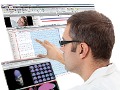
Operating in parallel since 2000, when Neuroscan acquired CURRY from Philips, the source reconstruction group in Hamburg, Germany has developed advanced algorithms for segmentation and source reconstruction that have led to the most sophisticated neuroimaging software package in the world — CURRY.
CURRY is used worldwide in a variety of research facilities, and in an increasing number of clinical operations, dealing primarily with epilepsy, where CURRY’s ability to integrate EEG and MEG with the CT, MRI, and other forms of image data provide an insight into the origin of the epileptic activity.
Neuroscan merged with Compumedics in 2003, and there was then a third R&D division, devoted primarily to the development of EEG amplifiers and software systems related to sleep disorders.
In recent years, there has been an inevitable and deliberate integration of the three overlapping R&D divisions, culminating in the CURRY Neuroimaging Suite, or CURRY 7. This suite of modular programs combines the years of software development for EEG acquisition and analysis from SCAN and the Compumedics acquisition software, with the image data analysis and source reconstruction from CURRY. CURRY 7 has been designed to acquire data not only with the Neuroscan amplifiers, but also with the Compumedics amplifiers. Data files from SCAN and from the Compumedics software are all read directly by CURRY 7.
CURRY 7 provides a solid foundation for years of future development in the research world, while also becoming more ensconced in clinical facilities. Because of its modular development, users need only obtain the components required for their current needs. The blend of R&D resources within Compumedics/Neuroscan ensures that the future will see even more functionality, as we continually strive to provide the best and most current capabilities for our users.
For additional details please contact sales@neuroscan.com or sales@compumedicsneuroscan.com
The post Research – Now and Into the Future appeared first on Compumedics Neuroscan.
Compumedics Neuroscan is dedicated to expanding knowledge and understanding of the human brain and nervous system through advanced technology.
Compumedics Neuroscan is a world-leading developer of research software for neurophysiology, neuroimaging, and neurodiagnostic systems. Neuroscan provides tools to increase understanding and improve treatment of this most complex and least understood system of the human body; the brain.
Neuroscan, founded in 1985, is the world’s leading provider of technologies for high-density EEG recordings, electro-magnetic source localization, multi-modal neuroimaging and enhancements to functional MRI. Neuroscan’s products are in use at over 1500 universities, corporate laboratories and national research institutes in approximately 40 countries.
Using the expertise acquired during the evolution of its high-level research products, Compumedics and Neuroscan have also developed clinical systems for EEG and EMG applications. Simple to use, yet employing Neuroscan’s sophisticated recording and signal processing techniques, the Neuroscan line of clinical products offer a high degree of value and performance.
In support of its customers, products and applications, Neuroscan also operates Neuromedical Supplies, a leading manufacturer and distributor of a wide range of accessory and disposable items used in both research and clinical neurology settings. Additionally, as a result of the merger with Compumedics Limited, a global supplier of clinical systems for polysomnography, neurology and cardiology, Neuromedical Supplies is now proud to also offer a complete range of high quality sensors and supplies for modern sleep laboratories.
The combined product range from Neuroscan and Compumedics is one of the largest in the industry. From compact 24-hour ambulatory EEG recorders for the hospital clinician to highly complex multimodal neuroimaging for large university research centers, this is the source.
Neuroscan and Compumedics — Partners in Neuroscience — Partners in Neurodiagnostics
The post About Neuroscan appeared first on Compumedics Neuroscan.
 Compumedics Neuroscan is pleased to announce a satellite symposium at the BIOMAG 2016 meeting in Seoul, Korea. The symposium “Comparison and combination MEG and EEG data” will be held October 2, 2016. More information is available on the website of BIOMAG.
Speakers:
In addition to this event, Compumedics is exhibiting at the BIOMAG meeting.
The post Satellite Symposium at BIOMAG 2016 appeared first on Compumedics Neuroscan.
Frequently Asked Questions about Scan
The post SCAN Frequently Asked Questions (FAQ) appeared first on Compumedics Neuroscan.
The documentation of CURRY 8 is available for download from our website (71Mb).
The contents of the ZIP archive is protected by a password. Please contact us to request the password for unpacking.
The post CURRY 8 PDF Documentation appeared first on Compumedics Neuroscan.
 Compumedics Neuroscan is pleased to announce CURRY 8, our premier tool for EEG/MEG signal processing and neuroimaging, has been released. It is now available for purchase. Please talk to your local representative in case you are interested in CURRY 8 or when you wish to upgrade you existing CURRY license.
The post CURRY 8 Released appeared first on Compumedics Neuroscan.
 The website has been updated with new information about electrode digitization. Compumedics Neuroscan supports a large variety of electrode digitization hardware. These include the NDI Krios, Spectra and Vicra devices as well as the Polhemus Fastrak and Patriot devices. The procedure of the electrode digitization is completely integrated in the CURRY Package and allows fast digitization of the electrode position with high accuracy.
The post Update: Electrode Digitizers appeared first on Compumedics Neuroscan.
We have uploaded a new video describing the key features of our Orion LifeSpan MEG System.
The post Update: Orion LifeSpan MEG appeared first on Compumedics Neuroscan.
 Compumedics Limited (ASX: CMP) (“Compumedics” or “Company”) is pleased to announce the confirmation of its first MEG contract to the world-renowned Barrow Neurological Institute (BNI), based in Phoenix, Arizona, USA.
The post Compumedics wins major multi-million dollar MEG brain imaging contract appeared first on Compumedics Neuroscan.
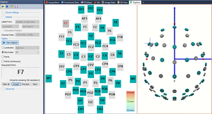
 Continuous helium-recycling minimizes the operating costs and maintenance requirements for magnetoencephalography. The Orion LifeSpan™ MEG System uses efficient helium-recycling with zero-loss.
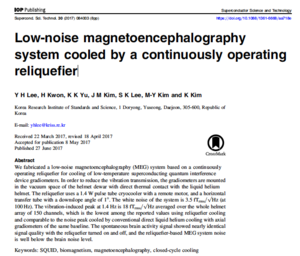
A recent publication by Lee et al. describes the helium-recycling system, the MEG system and the corresponding results.
The post Low-noise MEG by continuously operating reliquefier appeared first on Compumedics Neuroscan.
The documentation of Curry 7 is available for download from our website (55Mb).
The contents of the ZIP archive is protected by a password. Please contact us to request the password for unpacking.
The post Curry 7 PDF Documentation appeared first on Compumedics Neuroscan.
The post Orion LifeSpan MEG System overview appeared first on Compumedics Neuroscan.
We have set up a new website dedicated to our Orion LifeSpan MEG system. Please visit orionmeg.com to find out the latest about our unique MEG solution.
The post New website Orion LifeSpan MEG appeared first on Compumedics Neuroscan.
Compumedics Neuroscan is pleased to announce the successful installation and first phase commissioning of the Orion LifeSpan™ magnetoencephalography (MEG) at Barrow Neurological Institute (BNI) in Phoenix, Arizona, USA. Compumedics is also in the process of submitting its application for FDA 510(k) clearance, which will allow for clinical use of the MEG device, primarily for epilepsy and pre-surgical brain function mapping. This milestone marks the beginning of an exciting clinical and neuroscience research program planned at the prestigious neuroscience institute.
This successful installation marks a milestone for the MEG, with the Orion LifeSpan™ MEG being the first completely new design of a commercial MEG instrument to be delivered and installed in almost twenty years.
BNI, the world’s largest neurological disease treatment and research institution, is consistently ranked as one of the best neurosurgical training centers in the world. The Institute was founded in 1962 and has since grown to be one of the premier facilities in the world for neurology and neurosurgery, with more operative neurosurgical procedures undertaken at BNI than at any other USA institution.
Orion LifeSpan™ MEG technology has evolved from more than thirty years experience with magnetoencephalography (MEG) and electroencephalogram (EEG) technologies. Included are innovations in acquisition/analysis/visualisation software, highly sensitive magnetic field detectors and low-noise amplifier electronics, which have been developed at both the Korea Research Institute of Standards and Science (KRISS) and within Compumedics Neuroscan itself.
Ground-breaking features of the Orion LifeSpan™ MEG include advanced Superconducting Quantum Interference Device (SQUID) detectors for unparalleled sensitivity to brain signals; reduced operating cost from zero-loss helium reliquification with 24/7 operation; a fully integrated low-noise, high-density EEG monitoring system utilising the latest Compumedics/Neuroscan technology.
These hardware advancements are fully integrated with the state-of-the-art FDA-approved coregistration, neuroimage processing, and source estimation software known as CURRY – the world’s gold standard for clinical MEG/EEG and neuroscience research. Orion LifeSpan™ MEG also allows for a unique dual-helmet sensing system, with one side optimised for adult MEG recordings and the other for paediatrics. The exclusive pediatric capability will shortly be implemented at BNI, during the second and final installation phase.
